true

BWH Psychiatric Neuroimaging Faculty

Working in close collaboration with the Departments of Radiology, Medicine, and Neurology our faculty develop advanced imaging acquisition, analysis, and harmonization techniques to investigate disordered brain structure and function in psychiatric disorders as well as to develop new treatment strategies.

Overview

Psychiatric neuroimaging research at Brigham and Women’s Hospital integrates advanced imaging, computational methods, and clinical neuroscience to investigate brain structure, connectivity, and function in psychiatric and neurological disorders. Investigators employ multimodal approaches—including structural and functional MRI, diffusion imaging, and neurophysiologic methods—alongside machine learning and quantitative modeling to study conditions such as schizophrenia, mood disorders, OCD, and neurodevelopmental and stress-related disorders. Faculty work in close collaboration with colleagues in the Departments of Radiology, Neurology, and Medicine. The program emphasizes translational impact, using these tools to develop imaging biomarkers, improve neuromodulation targeting, and enable more precise, mechanism-based interventions.

Our Faculty

Martha E. Shenton PhD
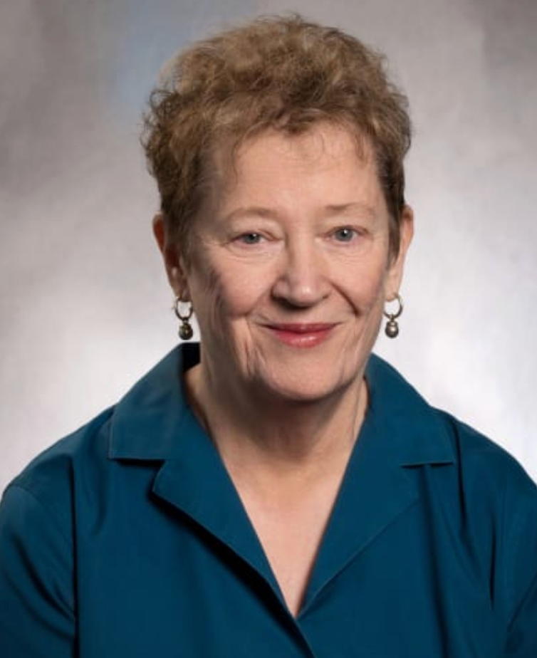
Dr. Shenton is the Director of the Psychiatry Neuroimaging Laboratory at BWH. Her research program applies advanced neuroimaging and image analysis methods to characterize structural and connectivity abnormalities underlying neuropsychiatric disorders, with a primary focus on schizophrenia. Using high-resolution MRI, diffusion tensor imaging, and novel computational approaches, her work has identified abnormalities in temporal lobe and limbic structures, as well as disrupted fronto-temporal connectivity, linking these findings to cognitive and language dysfunction. Her research extends to early-stage schizophrenia and related conditions to better understand disease onset and neurodevelopmental mechanisms, and incorporates techniques such as TMS to map brain–behavior relationships. In parallel, she has led major efforts using diffusion imaging and tractography to study white matter injury in traumatic brain injury and chronic traumatic encephalopathy, including large multi-site consortium work. Across these domains, her program aims to develop sensitive imaging markers that improve understanding of brain structure, connectivity, and function in clinical populations.
Martha Shenton is depicted

Marek Kubicki, MD, PhD

Dr. Kubicki’s research program focuses on developing and applying advanced neuroimaging methods to characterize structural and connectivity abnormalities underlying neuropsychiatric disorders, particularly schizophrenia. Using multimodal approaches—including structural MRI, diffusion tensor imaging, and related techniques—his work has been instrumental in identifying white matter abnormalities and disrupted fronto-temporal connectivity associated with psychosis and cognitive dysfunction. A central emphasis of his research is understanding how microstructural changes, including myelin alterations and neuroinflammation, contribute to disease onset, progression, and clinical symptoms. His program also integrates translational approaches across human, animal, and molecular studies to investigate mechanisms of psychosis, aging, and depression, and to develop high-resolution brain atlases and imaging biomarkers. Through this work, he aims to advance biologically grounded models of brain disorders and support the development of more targeted diagnostic and therapeutic strategies.
Marek Kubicki is depicted

Yogesh Rathi, PhD

Dr. Rathi’s research develops advanced computational and MRI techniques to better understand the structure and function of the living human brain. Drawing on tools from signal processing, statistics, control theory, and artificial intelligence, Dr. Rathi tackles challenging “inverse problems” in medical imaging—essentially, turning complex raw scanner data into clear, accurate pictures of brain tissue and connections.

Key innovations from his group include novel acquisition and reconstruction methods for faster diffusion MRI scans and ultra-high-resolution techniques that reveal fine details of brain microstructure (for example, properties of axons and other tissue features). A major focus is mapping brain connectivity using diffusion MRI tractography, alongside advanced analysis of functional data from fMRI, and EEG.

In parallel, Dr. Rathi’s team translates these tools into clinical applications. They develop precise tractography-based methods to improve targeting for neuromodulation therapies—including deep brain stimulation and transcranial magnetic stimulation—in conditions such as obsessive-compulsive disorder, Parkinson’s disease, and major depressive disorder. Recent work also emphasizes harmonizing multi-site MRI data and pushing the boundaries of mesoscale imaging to make advanced techniques more reliable and widely usable across ages and disorders. He is actively involved in developing statistical techniques for large-scale data analysis to understand the overlap and differences across a wide variety of brain disorders from a neural circuit perspective.

By bridging cutting-edge methodological innovation with direct clinical needs, Dr. Rathi’s program aims to enable more accurate, personalized, and data-driven interventions for neuropsychiatric and neurological conditions.
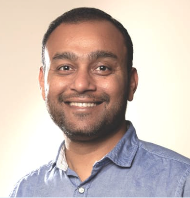

Yogesh Rathi is depicted

Laura M Holsen, PhD

The Holsen Lab conducts clinical neuroscience research focused on identifying aberrant activation patterns in eating, mood, and substance disorders. We use a multimodal approach (task-based fMRI, arterial spin labeling, and resting-state fMRI) to identify neural nodes and circuits in these conditions and collaborate with experts in neuromodulation and clincial assessment to inform the development of novel therapeutics.
Laura Holsen is depicted

Ofer Pasternak, PhD

Dr. Pasternak's research program develops advanced diffusion MRI methods to characterize brain microstructure and connectivity in neuropsychiatric and neurological disorders. He is widely recognized for introducing free-water imaging, a technique that separates extracellular water from tissue-specific diffusion signals, improving the sensitivity and specificity of diffusion MRI to detect pathology. A central focus of his work is using these methods to investigate white matter abnormalities, neuroinflammation, and microstructural changes associated with conditions such as schizophrenia, depression, and brain aging. His research also emphasizes multi-site harmonization and large-scale analysis of diffusion imaging data to improve reproducibility and enable population-level studies. Through this combination of methodological innovation and clinical application, his work aims to establish more precise imaging biomarkers of brain pathology and disease progression.
Ofer pasternak is depicted

Zora Kikinis, PhD

Dr. Zora Kikinis’s research program uses advanced neuroimaging to investigate the biological origins of mental disorders, with a focus on brain structure, connectivity, and their relationship to cognitive and behavioral dysfunction. Her current work centers on neuropsychiatric manifestations of long COVID (neuro-PASC), examining how cognitive deficits, fatigue, pro-inflammatory markers, viral factors, and alterations in white matter contribute to disability. Using multimodal MRI, particularly diffusion imaging, she aims to identify neural mechanisms underlying “brain fog” and related symptoms with significant public health impact. Her prior research has characterized white matter and gray matter abnormalities in schizophrenia and 22q11.2 deletion syndrome, linking early structural changes to neuropsychological deficits and risk for psychosis. Across a broad range of collaborative studies—including work in obesity, autonomic function, pain, traumatic brain injury, and women’s health—her program integrates imaging with clinical and biological data to advance mechanistic understanding and support the development of translational biomarkers.
Zora kikinis is depicted

Lipeng Ning, PhD

Dr. Lipeng Ning’s research program develops advanced MRI acquisition, reconstruction, and computational methods to characterize brain microstructure and network organization. His work focuses on multi-dimensional diffusion MRI and joint relaxation–diffusion imaging to improve the sensitivity and specificity of microstructural measurements, as well as novel algorithms for estimating tissue properties from complex imaging data. A central emphasis of his research is integrating diffusion and functional MRI to study brain network dynamics, applying concepts from control theory and information theory to understand causal relationships and system-level organization. In parallel, his program leverages machine learning and advanced signal processing techniques to address key challenges in neuroimaging, including data harmonization across scanners and scalable analysis of large datasets. His work also has a translational focus, applying these methods to guide and evaluate neuromodulation therapies such as transcranial magnetic stimulation and electroconvulsive therapy. Through this integration of methodological innovation and clinical application, his research aims to enable more precise characterization of brain circuits and improve intervention strategies in neuropsychiatric disorders.
Lipeng Ning is depicted

Suheyla Cetin Karayumak, PhD

Dr. Cetin-Karayumak's research program sits at the intersection of computer science, artificial intelligence, and medical image processing. Drawing on this computational foundation, she has spent the past several years developing advanced machine learning and image analysis methods for diffusion MRI, with a particular focus on multi-site data harmonization, white matter microstructure modeling, and tractography-based connectivity analysis. Her harmonization work has been validated through international benchmarking challenges and deployed across major multi-site neuroimaging consortia. Broadly, her research integrates methodological innovation in quantitative imaging with large-scale clinical applications spanning psychiatric and neurological populations, with the overarching goal of enabling robust, reproducible biomarker discovery across heterogeneous acquisition environments.
Suheyla Karayumak is depicted

Johanna Seitz-Holland, MD, PhD

Dr. Johanna Seitz-Holland’s research program applies advanced neuroimaging and statistical modeling approaches to understand how brain structure relates to cognition, physical health, and clinical symptoms in psychiatric disorders, with a primary focus on psychosis. Using multimodal MRI alongside clinical, cognitive, and biological data, her work investigates how factors such as age, sex, and environmental influences shape brain organization and disease risk. A central emphasis of her research is identifying interactions between brain structure and systemic health, including emerging work integrating neuroimaging with proteomics to study cellular aging and brain health in psychosis. Her program also examines modifiable risk factors—such as sleep and social behavior—and their relationship to mental illness. Through this integrative and translational approach, her work aims to advance biologically grounded models of psychiatric disorders and support the development of more precise biomarkers and interventions.
joanna seitz-holland is depicted

Joseph J. Taylor, MD, PhD

The Interventional Psychiatry Research Program (IPRP) aims to develop and test procedural treatments for individuals with psychiatric illnesses who have not benefited from conventional psychotherapy and medications. Most of our work involves deriving, validating, and testing circuit-based treatment targets for brain stimulation or neurosurgical ablation. The IPRP works in close collaboration with the Center for Brain Circuit Therapeutics, a Division of the Neuropsychiatry and Interventional Psychiatry program.
joseph taylor is depicted