Brigham Research Imaging Core (BRIC)
Email: klnolan@bwh.harvard.edu
Call: 617-525-8945
Overview
The BWH Research Imaging Core (BRIC) provides a comprehensive research imaging service to meet the needs of investigators and research subjects using imaging facilities at Brigham and Women’s Hospital (BWH). A unique feature of BRIC is the complete anonymity of research subjects. Research image scheduling, image acquisition and image storage are all kept separate from BWH clinical Radiology systems. The BRIC provides the administrative infrastructure, customer service architecture and institutional support to promote investigative applications of imaging technologies.
Services and Equipment
BWH Research Imaging Core (BRIC) Services
BRIC offers image acquisition and storage services using state-of-the-art equipment at BWH. A list of available imaging equipment can be found further below.
BRIC also offers image upload and storage of images obtained outside of BWH and imported into the BWH research PACS system.
Imaging Study Initiation/Scheduling
A significant component of the BRIC’s infrastructure is information technology in the form of a Research PACS that mirrors the Clinical PACS in all functions and more. The research subject’s data and information are kept separate from the clinical/MRN system at all stages of the project—from scheduling to image acquisition and data management. Study subject screening and scheduling as well as image protocol and resultant data interpretation are provided in compliance with IRB protocols. To begin a new project please complete a BRIC Intake form and email it to Karen Nolan at klnolan@bwh.harvard.edu.
Safety Reads
All subjects have a basic minimum set of images acquired in every imaging study per their research protocol. In all cases, a board certified/board eligible radiologist interprets this basic set of images, evaluating for clinically significant incidental findings. Radiology interpretation is limited to diagnostic sequence, not the Imaging research sequences acquired unless further radiology research assessment is contracted. Subject identity and/or link to insurance information is kept private. The cost of the safety read is included in BRIC scanning costs.
See link to BRIC Policy on Radiologist Interpretation of Images
Image Storage – BWH and non-BWH Images
Image storage is provided through the research PACS system, including storage of research images from non-BWH institutions, and uploading of images from the BWH clinical PACS. There is a dedicated fee for importing and storing images from outside BWH and from the BWH clinical PACS.
Image Acquisition Experts
MRI and CT technologists are available during clinical scanning hours (and off-hours on a case by case basis) and their services are included in the scanning cost. Exceptions are for the small animal imaging scanners. We offer training for investigators to operate the Bruker In-Vivo Extreme II Optical/X-ray scanner and the Bruker Skyscan1176 microCT scanner. For the Bruker microMRI scanners we can provide a research specialist for a fee, if the investigator does not have previous MRI training.
BWH Research Imaging Core (BRIC) Pricing
Prices for our services are reviewed annually and may change each year in response to market conditions, operational costs, and other economic factors. To view a PDF with the BWH Research Imaging Core User Fees for FY26, click here.
Application Forms
- Image Acquisition and Storage - Human Imaging
- Image Acquisition and Storage - Animal Imaging
- Image Acquisition and Storage – Phantom Imaging for Internal Mass General Brigham Researchers
- Image Acquisition and Storage – Phantom Imaging for Non-Mass General Brigham Researchers
- Image Upload to Research PACS
- Special Acquisition Services
GE 3T MAGNUS Research Only MRI Scanner
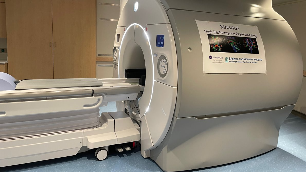
MRI Equipment and Locations
GE 3T MAGNUS: 221 Longwood Ave. Ground Floor
GE 3T-Short Bore: 221 Longwood Ave. Ground Floor
Siemens 3T Skyra: 221 Longwood Ave. Ground Floor
Siemens 3T Verio (Bay 2): ASB2 L1
GE Discovery 750W 3T (Bay 3): ASB2 L1
Siemens 3T Trio (Lee Bell): CWN 2nd Floor
Siemens 3T Trio: Shapiro Building L2
Siemens 3T Prisma (2): Building for Transformative Medicine L2
Siemens 3T Skyra: Building for Transformative Medicine L2
Siemens 1.5T Aera: Building for Transformative Medicine L2
Siemens 7T Terra: Building for Transformative Medicine L2
CT Equipment and Locations
Toshiba Aquilion One 320: Shapiro Building L2
Siemens Definition Dual Source/Dual Energy: Shapiro Building L2
Siemens Definition Dual Source/Dual Energy: Building for Transformative Medicine L2
Small Animal Imaging Equipment and Locations
Bruker 7T with Cryoprobes: 221 Longwood Ave. Ground Floor
Bruker Biospec 3T: Building for Transformative Medicine, 11th Floor
Bruker SkyScan 1176 mCT: Building for Transformative Medicine, 11th Floor
Bruker In-Vivo Extreme II Optical/X-ray: Building for Transformative Medicine, 11th Floor
Sedecal SuperArgus 4R: Building for Transformative Medicine, 11th Floor
Small Animal Imaging Laboratory (SAIL) for pre-clinical imaging
SAIL is comprised of two facilities conveniently located within the Longwood Medical Area: SAIL Satellite at 221 Longwood Ave and Imaging Crescent at 60 Fenwood Rd (11th floor of HBTM). Its proximity to multiple medical and research institutions, as well as Harvard Medical School, allows for a more naturally collaborative environment by providing investigators with a comprehensive, translational research infrastructure.
SAIL includes access to multiple vivariums and fully equipped preparatory areas for pre- and post-imaging procedures and enables longitudinal studies. Investigators also have access to nearby housing facilities for larger models. These housing facilities are connected to the BWMRC via an underground hallway, which avoids the need to hire transport for the larger models.
7.0T Bruker BioSpec® USR magnet for high spatial resolution imaging of small models.
3T Bruker Biospec® featuring a motorized transport system; includes multiple RF coils.
3.0T Siemens Skyra wide-bore magnet available for imaging medium to large models.
Magnetic Resonance Imaging (MRI) is an essential research tool for the non-invasive study of structure and function. The ability to use this technology in pre-clinical models allows translational studies that are crucial to elucidating a variety of disease mechanisms and to developing novel treatment approaches. From the study of brain function in neuropsychiatric disease, to the measurement of pulmonary function, to the introduction of new, minimally invasive image-guided treatment approaches, the use of novel MRI research methods is revolutionizing biomedical science.
The Bruker Skyscan1176 provides for ultra-low X-ray dose in vivo scans for longitudinal studies with minimal radiation effects. In vivo imaging can be performed at nominal resolutions of 9, 18 or 35 microns with a maximum FOV of 68mm x 200mm. With the ability to alter beam energy, exposure time, and filtering this system is excellent for both soft tissue and bone imaging. One of the applications of the scanner is bone studies where the morphometric parameters such as bone mineral density, bone volume ratio and trabecular thickness can be extracted from the images. It can monitor bone architecture changes over time, evaluating possible healing treatments or regenerations and understanding skeletal development or deformity progress like rheumatoid arthritis.
Micro CT can also be used for various lung-disease studies including lung fibrosis. The low radiation dose as well as the ability to synchronize scans with respiratory cycles provides excellent in vivo longitudinal lung imaging.
BRIC Policies and Procedures, FAQ’s
Is the BWH Research Imaging Core appropriate for all research projects that include imaging?
No, the BRIC is not appropriate for research projects or clinical trials where the imaging exams need to be included in the patient/subject's medical record. All imaging exams that are done through the BRIC are stored in a separate research PACS system which is not connected to Epic or the subject’s chart.
Why should I include the Imaging Core in my extramural funding budget (NIH grant, industry sponsored clinical study) instead of budgeting imaging as patient care expenses?
When you are budgeting for study “subjects” the Imaging Core will provide more flexibility to the investigator. The Imaging core is not tied to the predefined scanning that is used for patient care and can be customized to the investigator’s study design/protocol. Costs are based on scan time and can be scheduled into multiple timeslots. Also, the direct cost of Imaging Core scans is generally lower priced than the patient care equivalent.
When budgeting for the Imaging Core, the expense is budgeted as a direct cost in the “other” section of the budget and comes fully loaded with IDC. If for any reason you had to switch from “subjects” to “patients” the rebudgeting would work in the investigators favor. However, if the line item had been originally budgeted as patient care, and the investigator decides that they would like to use the Imaging Core, the rebudgeting works against the investigator. Patient care is not awarded with IDC, and the imaging core expenses would have IDC associated with them.
When would I not want to include the Imaging Core in my extramural funding budget?
In the case where you are submitting a NIH proposal that has a cap on total cost, it may seem more expensive to use the imaging core, because the scans which are generally lower priced than a patient care equivalent, do bear overhead, making the total cost of the scan more expensive
On Industry Sponsored Clinical Trials should I use Patient Care Expense or Imaging Core?
Since the corporate sponsored Clinical Trials have a Total Direct Cost base, the determining factor will be the subject population. If subjects are “patients” and the information needs to be recorded in their medical record, then patient care charges will be applied. If the trial requires healthy volunteers or “subjects” that are not under care requiring the information to be on their medical record, the Imaging Core may be preferable. In most cases Industry Sponsored Clinical Trials should be budgeted as Patient Care. Industry Trials could have a combination of patient care and Imaging Core.
Will MRI scans completed using the Imaging Core still be read by a clinical Radiologist and will this generate a clinical report?
There will be a clinical Radiologist read of all scans completed using the Imaging Core, this is included in the pricing. No clinical report will be included in the subject's medical record (if you need this, then you should not be using the Core). If the Radiologist sees a significant finding, they will contact the PI and it will be up to the PI to get permission from the subject to contact their PCP to report the finding. The PI will need to help the subject find a PCP if they don't have one. At the request of the Principal Investigator and with permission from the participant, images can be transferred to their clinical chart in Epic if follow-up by an MGB physician is planned.
How will the new research record numbers be assigned to study subjects?
We need to ensure that codes are consistent with the codes/identifiers assigned to the study subjects in our research project. The modality for patient anonymization will need to be understood.
The subject research record number (RRN) will be generated from the BRIC REDCap scheduling system and assigned to the subject to ensure anonymity. The RRN will get assigned when the patient is registered in REDCap. If a research subject is followed longitudinally in a study, each time they are scheduled in REDCap they will be scheduled with the same RRN. Each new exam will have a different accession number assigned to it but the RRN will remain the same (just as an MRN would).
The RRN is not the same as the study subject ID assigned in each study, the RRN will be used to schedule the subject in the REDCap system. It will be up to the research coordinator to keep a confidential internal record of subject names, study IDs and RRNs according to IRB standards.
Which MRI Scanners are currently included in the Research Imaging Core?
Please refer to the equipment list above